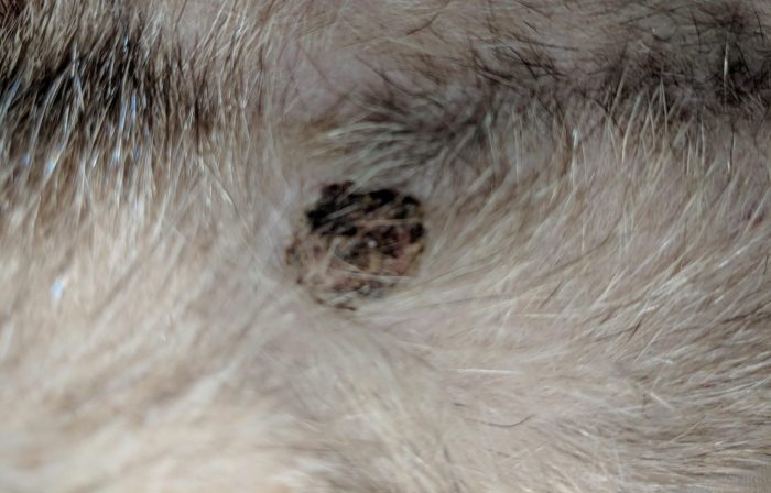
Ferret mast cell tumor picture provides a comprehensive overview of this prevalent health concern in ferrets, offering insights into its clinical presentation, diagnostic techniques, treatment options, and prognosis. This article delves into the intricacies of mast cell tumors in ferrets, empowering owners with the knowledge to make informed decisions regarding their beloved pets’ health.
Mast cell tumors are a common type of skin cancer in ferrets, characterized by their aggressive behavior and potential for metastasis. Understanding the various aspects of this disease is crucial for ensuring the best possible outcomes for affected ferrets.
Mast Cell Tumors in Ferrets

Mast cell tumors (MCTs) are a common type of cancer in ferrets, accounting for approximately 10-25% of all tumors diagnosed in this species. They are typically malignant and can occur in any part of the body, although the most common sites are the skin, gastrointestinal tract, and spleen.MCTs
in ferrets can vary in appearance and clinical presentation, depending on their location and stage of development. Some common signs and symptoms include:
- Skin lesions: MCTs on the skin may appear as raised, red, or purple bumps or nodules. They can be firm or soft and may ulcerate or bleed.
- Gastrointestinal signs: MCTs in the gastrointestinal tract can cause vomiting, diarrhea, abdominal pain, and weight loss.
- Splenic involvement: MCTs in the spleen can lead to an enlarged spleen, which can cause abdominal discomfort and breathing difficulties.
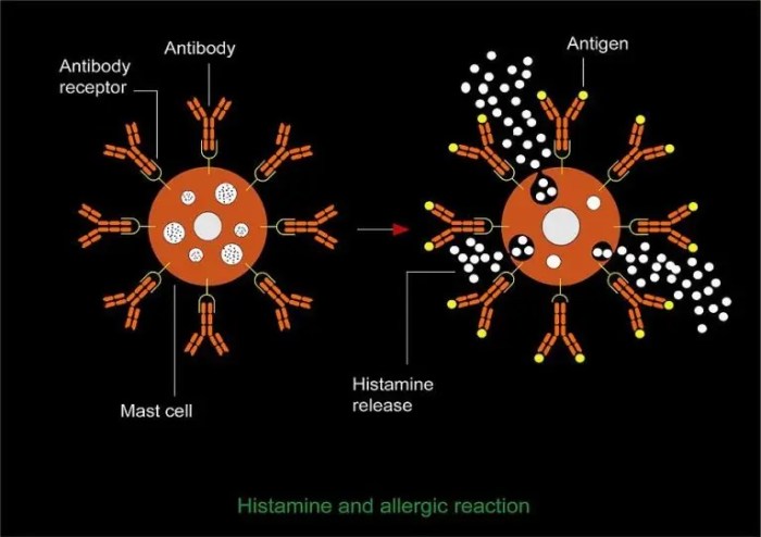
- Systemic signs: In advanced cases, MCTs can release histamine and other inflammatory mediators into the bloodstream, causing systemic signs such as anaphylaxis, hypotension, and shock.
There are several different types of MCTs that can occur in ferrets, each with its own characteristics:
- Cutaneous MCTs: These are the most common type of MCT in ferrets and typically occur on the skin. They can be solitary or multiple and may range in size from small bumps to large masses.
- Gastrointestinal MCTs: These MCTs occur in the gastrointestinal tract and can cause a variety of clinical signs, including vomiting, diarrhea, abdominal pain, and weight loss.
- Splenic MCTs: These MCTs occur in the spleen and can lead to an enlarged spleen, which can cause abdominal discomfort and breathing difficulties.
- Disseminated MCTs: These MCTs have spread to multiple organs or tissues in the body and are often associated with a poor prognosis.
Diagnostic Techniques for Mast Cell Tumors in Ferrets

Confirming the presence of mast cell tumors in ferrets requires a comprehensive diagnostic approach. Various techniques are employed to accurately identify and characterize these tumors.
Cytology and Histopathology
Cytology and histopathology are essential diagnostic techniques for mast cell tumors in ferrets. Cytology involves examining cells obtained from a fine-needle aspirate or skin scraping under a microscope. This technique provides a rapid and minimally invasive means of assessing the cellular characteristics of the tumor, including the presence of mast cells and their degranulation status.
Histopathology, on the other hand, involves examining tissue samples obtained through a biopsy under a microscope. This technique allows for a more detailed evaluation of the tumor’s architecture, cell type, and degree of differentiation. Histopathology is considered the gold standard for diagnosing mast cell tumors and determining their grade and prognosis.
Imaging Techniques
Imaging techniques, such as X-rays and ultrasound, play a valuable role in evaluating mast cell tumors in ferrets. X-rays can help identify skeletal involvement or distant metastases, while ultrasound can assess the size, location, and internal structure of the tumor.
These imaging techniques provide important information for staging the tumor and guiding treatment decisions.
Differentiating Mast Cell Tumors from Other Skin Conditions
Differentiating mast cell tumors from other skin conditions in ferrets is crucial to ensure appropriate treatment. Mast cell tumors can mimic various skin lesions, including allergies, abscesses, and other types of tumors. A thorough clinical examination, combined with cytology or histopathology, is essential for accurate diagnosis.
In the realm of veterinary medicine, ferret mast cell tumors often captivate the attention of researchers. While delving into the intricacies of these tumors, one may stumble upon the existence of the compound K2O2. The compound K2O2 , possessing intriguing properties, has been the subject of numerous studies.
Returning to the topic of ferret mast cell tumors, their complex nature continues to unravel as scientists explore novel avenues for diagnosis and treatment.
By utilizing a combination of diagnostic techniques, veterinarians can accurately identify and characterize mast cell tumors in ferrets, enabling them to develop optimal treatment plans and provide the best possible care for their patients.
Treatment Options for Mast Cell Tumors in Ferrets: Ferret Mast Cell Tumor Picture

Mast cell tumors in ferrets can be treated with a variety of modalities, including surgery, radiation therapy, and chemotherapy. The best treatment option for a particular ferret will depend on the size, location, and grade of the tumor, as well as the ferret’s overall health.
Surgery
Surgery is the most common treatment for mast cell tumors in ferrets. The goal of surgery is to remove the entire tumor, including any surrounding tissue that may be affected by the tumor. The type of surgery performed will depend on the location of the tumor.
In some cases, it may be possible to remove the tumor with a simple excision. In other cases, more extensive surgery may be necessary, such as a mastectomy (removal of the mammary gland) or a limb amputation.
Radiation Therapy
Radiation therapy is a treatment option for mast cell tumors that are located in areas that are difficult to surgically remove, such as the head or neck. Radiation therapy uses high-energy beams of radiation to kill cancer cells. Radiation therapy can be given externally, using a machine that delivers radiation to the tumor from outside the body, or internally, using radioactive implants that are placed directly into the tumor.
Chemotherapy, Ferret mast cell tumor picture
Chemotherapy is a treatment option for mast cell tumors that have spread to other parts of the body. Chemotherapy uses drugs to kill cancer cells. Chemotherapy can be given orally, intravenously, or through a port that is surgically placed under the skin.
Prognosis and Management of Mast Cell Tumors in Ferrets

Ferrets with mast cell tumors have a variable prognosis, influenced by factors such as tumor size, location, and grade. Smaller, well-differentiated tumors located in easily accessible areas tend to have a better prognosis than larger, poorly differentiated tumors located in internal organs.Regular
monitoring and follow-up care are crucial after treatment for mast cell tumors in ferrets. This includes physical examinations, blood tests, and imaging studies to assess tumor response and detect any recurrence or metastasis.
Managing Potential Complications
Ferrets with mast cell tumors may experience various complications, including:
Anaphylaxis
A severe allergic reaction that can occur due to the release of histamine and other inflammatory mediators from mast cells. Symptoms include difficulty breathing, swelling, and vomiting.
Gastrointestinal problems
Mast cell tumors in the gastrointestinal tract can cause vomiting, diarrhea, and abdominal pain.
Skin problems
Mast cell tumors on the skin can cause itching, redness, and swelling.
Bone marrow suppression
Mast cell tumors can release substances that suppress bone marrow function, leading to anemia, neutropenia, and thrombocytopenia.These complications require prompt veterinary attention and may necessitate additional treatment measures, such as antihistamines, steroids, antibiotics, or blood transfusions.
Question Bank
What are the most common symptoms of mast cell tumors in ferrets?
The most common symptoms include skin lesions, itching, gastrointestinal upset, and respiratory problems.
How are mast cell tumors diagnosed in ferrets?
Diagnosis typically involves a combination of cytology, histopathology, and imaging techniques.
What are the treatment options for mast cell tumors in ferrets?
Treatment options include surgery, radiation therapy, and chemotherapy.
What is the prognosis for ferrets with mast cell tumors?
The prognosis depends on factors such as tumor size, location, and grade, but early detection and treatment can improve outcomes.